Atomic Force Microscope: Principle, Advantages & More
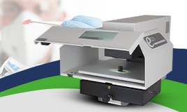
The Jupiter XR AFM is the world’s first large-sample atomic force microscope that offers high-speed imaging along with extended range in a single scanner. But before you learn about this product which is available at our BCL platform, make sure to understand what exactly is Atomic Force Microscope.
It is a type of scanning probe microscope that primarily functions to measure properties such as magnetism, height, friction. The resolution is performed in a nanometre unit that is more precise and effective in comparison to the optical diffraction limit.
This microscopy technique uses a probe for the measurement and collection of data which involves touching the surface required for probing. An image is developed when the instrument raster-scans the probe upon the sample’s section, measuring its local properties parallelly.
Keep reading till the end to find out more about atomic force microscopes.
Atomic Force Microscope
This microscopy solution also has piezoelectric elements that are basically electrical charges which accumulate over specific solid materials such as biological proteins, DNA, crystal, etc and therefore allow tiny accurate movement when scanning upon an electric program.
The atomic force microscope was invented in 1982 right after the foundation of scanning tunnelling microscopes two years before. However, it was only in 1986 that it was used for experiments and was commercialised for the market in 1989.
The Fundamental Principle of Atomic Force Microscope
This particular microscopy technique works on the principle of measuring intermolecular forces and examines atoms by making use of probed surfaces of the sample in nanoscale. Its function relies on 3 main working principles that are – surface sensing, detection and imaging.
- Firstly, the microscope performs surface sensing with the use of a cantilever which is an element made from rigid block such as a plate or beam which is attached to the end of support. The cantilever has a sharp tip that works to scan over the sample surface with the help of producing an attractive force between the tip and the surface when it is drawn closer to the surface of the sample. Finally, when it is drawn extremely close and makes contact with the surface, a repulsive force produces the cantilever to avert from the sample surface.
- When the cantilever deflects away from the sample surface, there is an alteration in the reflection beam’s direction and aids in detecting the aversion by a laser beam. The use of a positive-sensitive photo-diode (PSPD) monitors these deflection alterations and accurately records them.
- The third working principle is taking the image of surface topography of the specimen by force. This happens by scanning the cantilever over an area of interest. Based on how low or raised the sample surface is, the determination of the deflection beam is carried out which is again recorded by the PSPD.
Advantages of Using Atomic Force Microscope
- Sample preparation is done easily for the purpose of observation.
- Enabled with 3D imaging capabilities.
- Accuracy in sample size measurement.
- It can be used in air, vacuums and liquids.
- It can be used for quantifying the sample’s surface roughness.
- Ideal for usage in dynamic environments.
Investing on Jupiter XR AFM can further amplify the range of benefits that a standard atomic force microscope offers.


